What Is Karyotyping?
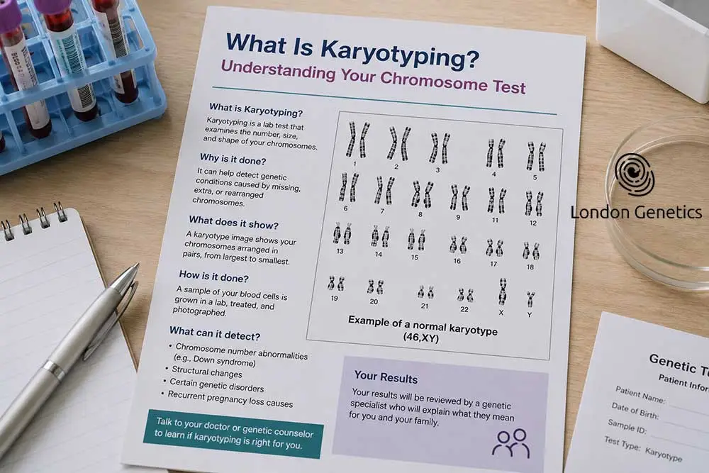
Karyotyping is a laboratory test that examines the number, size, and structure of a person’s chromosomes, the thread-like structures within our cells that carry our genetic information. Humans typically have 46 chromosomes, arranged in 23 pairs, and karyotyping produces a visual map of these chromosomes, allowing clinicians to check whether everything is present, correctly formed, and properly arranged.
The test works by taking a small sample, usually blood, though occasionally other tissue types, and growing the cells in a laboratory until they reach a stage of division where chromosomes become visible under a microscope. These chromosomes are then photographed, sorted by size and shape, and arranged into pairs, creating what’s known as a karyogram, the image most people picture when they think of karyotyping.
What makes this test so valuable is its ability to reveal chromosomal abnormalities that wouldn’t be detectable through standard blood tests or physical examination alone. Whether it’s an extra chromosome, a missing one, or a structural rearrangement, karyotyping provides clear, visual evidence of what’s happening at a chromosomal level, forming the basis for a confirmed diagnosis in many genetic conditions.
Though newer, more detailed genetic technologies now exist, karyotyping remains a well-established, reliable, and widely used starting point in genetic investigation across the NHS and private clinics alike.
How Is a Karyotype Test Carried Out?
Understanding what actually happens during a karyotype test can make the process feel far less daunting, whether you’re having one yourself or a family member is being tested.
The process usually begins with a simple blood sample, taken in much the same way as any routine blood test. In certain circumstances, such as prenatal testing, the sample may instead come from amniotic fluid or placental tissue, collected via amniocentesis or chorionic villus sampling.
Once in the laboratory, the sample’s cells are placed in a special growth medium and left to divide over several days. The dividing cells are then treated with a chemical that stops division at the right moment, before being stained to reveal distinctive banding patterns unique to each chromosome, and high-resolution photographs are taken under a microscope.
Using specialist software, the photographed chromosomes are digitally arranged into pairs, ordered by size, creating the completed karyogram. A trained clinical scientist then carefully examines it, checking for the correct number of chromosomes and any structural abnormalities, before compiling a formal report for your doctor or genetic counsellor to discuss with you.
From sample to result, a karyotype test typically takes one to three weeks, depending on the laboratory and the reason for testing, so a little patience is usually needed while the analysis is carried out thoroughly.
What Does a Normal Karyotype Look Like?
Once you understand what a karyotype actually shows, it becomes much easier to make sense of your own results, or those of a family member.
The Standard Chromosome Count
A typical human karyotype consists of 46 chromosomes, arranged into 23 matching pairs. Twenty-two of these pairs, known as autosomes, are identical in both males and females, while the 23rd pair determines biological sex.
Sex Chromosomes
In a normal female karyotype, the 23rd pair consists of two X chromosomes, written as 46,XX. In a normal male karyotype, this pair consists of one X and one Y chromosome, written as 46,XY. This shorthand notation is simply a standard way of summarising the total chromosome count alongside the sex chromosomes present.
Size and Structure
In a normal result, each chromosome pair matches in size, shape, and banding pattern, with no missing, extra, or rearranged material. Chromosomes are numbered 1 to 22 according to size, from largest to smallest, followed by the sex chromosomes.
What “Normal” Actually Confirms
A normal karyotype result confirms that no chromosomal abnormality was detected at the resolution the test can identify. It’s worth noting that karyotyping examines chromosomes as a whole, so it wouldn’t detect very small genetic changes within a single gene, that’s where more detailed tests, such as microarray analysis or sequencing, come in.
Seeing “46,XX” or “46,XY, normal karyotype” written on a report is generally reassuring, though your genetic counsellor or doctor will always explain the result within the wider context of why the test was carried out in the first place.
What Can Karyotyping Detect?
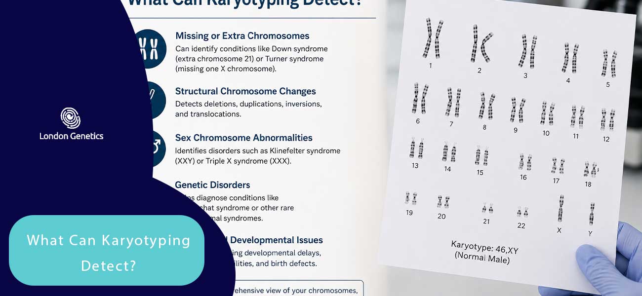
Karyotyping is particularly effective at identifying chromosomal abnormalities, changes in the number or structure of chromosomes, some of which are responsible for well-known common genetic disorders.
Extra or Missing Chromosomes
One of the clearest applications of karyotyping is detecting an extra or missing chromosome. Down syndrome, for example, occurs when there’s an extra copy of chromosome 21, resulting in 47 chromosomes rather than the usual 46, a finding karyotyping identifies clearly and reliably.
Sex Chromosome Variations
Karyotyping can also reveal differences in the number of sex chromosomes. Turner syndrome, where a female has only one X chromosome instead of two, and Klinefelter syndrome, where a male has an additional X chromosome, are both readily identified through this test.
Structural Chromosomal Changes
Beyond simple counting, karyotyping can detect structural abnormalities, including translocations (where part of one chromosome attaches to another), deletions (missing sections), and duplications (repeated sections), all of which can have significant health implications.
Prenatal and Fertility-Related Findings
In pregnancy, karyotyping helps identify chromosomal conditions in a developing baby, while in fertility investigations, it can uncover chromosomal factors contributing to recurrent miscarriage or difficulty conceiving.
What Karyotyping Cannot Detect
It’s equally important to understand the test’s limits. Karyotyping identifies changes visible at a whole-chromosome level, but it cannot detect smaller genetic mutations within individual genes, such as those responsible for cystic fibrosis or sickle cell anaemia. For these, more detailed genetic testing is required.
By clarifying which conditions fall within its scope, karyotyping plays a foundational role in the wider diagnostic picture of many common genetic disorders, often serving as the first, essential step towards a clear diagnosis.
When Is Karyotyping Recommended?
Karyotyping isn’t a routine test carried out on everyone; it’s typically recommended in specific circumstances where a chromosomal cause is genuinely suspected.
Your doctor or genetic counsellor will always explain exactly why karyotyping is being recommended in your specific situation, ensuring you understand what the test is looking for before proceeding.
Understanding Your Karyotype Results
Interpreting the findings of a chromosome analysis requires a structured, expert evaluation. When undergoing karyotyping, the resulting report—known as a karyogram—provides a comprehensive visual map and formal description of an individual’s chromosomal arrangement. Understanding how these results are structured helps patients and clinicians make informed decisions regarding diagnosis, management, and family planning.
1. The Standard Karyotype Nomenclature
A laboratory report following karyotyping uses a standardized international system of shorthand notation (ISCN) to describe chromosome composition. This formula typically consists of three primary elements:
-
Total Number of Chromosomes: The overall count observed per cell (typically 46 in a neurotypical profile).
-
Sex Chromosome Complement: The combination of sex chromosomes present (XX for females, XY for males).
-
Observed Abnormalities: Any structural or numerical variations identified during analysis.
Example Notations:
46,XX– Normal female karyotype.
46,XY– Normal male karyotype.
47,XY,+21– Male karyotype indicating Down’s syndrome (an extra copy of chromosome 21).
2. Differentiating Normal vs. Abnormal Findings
When evaluating karyotyping results, clinicians categorize findings based on whether structural or numerical variations are detected:
Normal Results
A normal result confirms that all 23 pairs of chromosomes are present, structurally intact, and showing expected banding patterns under microscopic examination. This rules out large-scale chromosomal rearrangements or major copy number variations.
Numerical Abnormalities (Aneuploidy)
An abnormal result may indicate missing or extra chromosomes:
-
Trisomy: The presence of three copies of a specific chromosome instead of a pair (e.g., Trisomy 18 or Edwards syndrome).
-
Monosomy: The absence of one chromosome from a pair (e.g., Turner syndrome, designated as
45,X).
Structural Abnormalities
Karyotyping can also identify structural changes where the total chromosome count remains 46, but the material within them has been altered:
-
Translocations: Portions of two different chromosomes have swapped places (balanced or unbalanced).
-
Deletions or Insertions: Segments of a chromosome are missing or duplicated.
-
Inversions: A chromosomal segment has detached, inverted 180 degrees, and reattached.
3. Mosaicism: Mixed Cellular Lineages
In some instances, karyotyping reveals a mosaic pattern—a condition where an individual possesses two or more genetically distinct cell populations derived from a single zygote. For instance, a report reading 46,XX / 47,XX,+21 indicates that some cells display a normal female profile while others carry an extra chromosome 21. The clinical impact of mosaicism often depends on the proportion of altered cells present in body tissues.
4. Next Steps Following Your Results
Receiving your karyotyping report is often one stage in a broader diagnostic process:
-
Post-Test Genetic Counselling: Whether results are normal or abnormal, consultation with a registered clinical geneticist or genetic counsellor is essential to contextualize findings.
-
Further Genomic Testing: Because traditional karyotyping focuses on macroscopic chromosome structures (typically resolving changes larger than 5–10 megabases), further high-resolution analysis—such as Chromosomal Microarray Analysis (CMA) or Whole Genome Sequencing—may be recommended if subtle microdeletions are suspected.
Summary: Key Takeaways on Karyotyping
Karyotyping is a foundational laboratory technique that provides a comprehensive bird’s-eye view of an individual’s genome by mapping and pairing all 23 sets of human chromosomes. It remains an essential diagnostic standard in modern clinical genetics.
By evaluating overall chromosomal structure and numerical arrangements, karyotyping bridges the gap between clinical symptoms and underlying genetic drivers. Understanding its diagnostic scope ensures families and clinicians select the most effective analytical pathway.