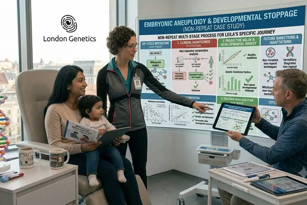
Defining Embryonic Aneuploidy: The Microscopic Counting Error
The Fundamentals of Chromosomal Architecture
To truly comprehend the complexities of early pregnancy complications, one must first look at the very foundation of cellular biology. Every typical human life begins with a precise genetic blueprint consisting of 46 chromosomes, neatly arranged into 23 pairs. Half of this genetic package is inherited from the egg, and the other half from the sperm. When a conception occurs with the exact required number of chromosomes, it is referred to as a euploid embryo, which possesses the standard biological structural code needed to develop into a healthy baby.
The Mechanism of a Cellular Miscount
The term embryonic aneuploidy refers to a specific type of genetic mismatch where this delicate numerical balance is disrupted. Instead of inheriting the standard 46 chromosomes, the developing embryo ends up with a missing or an extra chromosome:
-
Trisomy (An Extra Chromosome): This occurs when an embryo inherits three copies of a specific chromosome instead of the usual pair. A well-known clinical example of a live-birth trisomy is Down’s syndrome (Trisomy 21).
-
Monosomy (A Missing Chromosome): This happens when an embryo is missing a vital chromosome from its pair, leaving it with only 45 chromosomes in total.
-
The Point of Origin: This microscopic counting error usually takes place during meiosis—the specialized cell division that creates eggs and sperm. If the chromosome pairs fail to separate equally, the resulting reproductive cell carries an incorrect amount of DNA from the very beginning.
The Immediate Dynamic in Early Development
Following fertilisation, the cells begin to divide at an astonishing pace. If embryonic aneuploidy is present, every single subsequent cell copied from that initial blueprint will inherit the exact same structural flaw. While the embryo may appear structurally normal under a standard laboratory microscope during the first few days of division, the underlying genetic code is profoundly compromised. As we explore the broader context of reproductive health, understanding these microscopic variations becomes essential, particularly when evaluating how structural cellular vulnerabilities naturally shift alongside Advanced Maternal Age Genetic Risks.
The Biological Mechanics: How Chromosomal Abnormalities Halt Development
When a developing pregnancy carries an incorrect number of chromosomes, the impact on early cellular differentiation is catastrophic. In the initial weeks, the embryo relies on a perfectly balanced set of genetic instructions to orchestrate the formation of vital organs and complex bodily systems.
The underlying philosophy of modern reproductive science and predictive embryology lies in replacing reactive selection methods with strategic biomolecular foresight. Analysing how individual bioenergetic profiles and high-risk chromosomal variants interact with the maternal environment allows clinical teams to eliminate outdated trial-and-error transfer protocols. Mapping these specific pathogenic and architectural traits transforms abstract sequencing into an unshakeable, personalised blueprint for long-term health maintenance and successful pregnancy survival.
The Silent Escalation: Cellular Vulnerability Over Time
The Biological Reality of Oocyte Ageing
Unlike the paternal system, which continuously produces fresh reproductive cells throughout adulthood, the maternal biological clock operates on a strictly finite timeline. A woman is born with her entire lifetime supply of eggs, which remain in a state of suspended animation within the ovaries for decades. As time progresses, these delicate cells are exposed to natural metabolic wear and tear. This prolonged waiting period introduces a silent escalation in cellular vulnerability, directly increasing the statistical likelihood of embryonic aneuploidy once these eggs are fertilised.
The Mechanics of Meiotic Nondisjunction
When the body finally signals an egg to mature and prepare for potential fertilisation, the cell must complete a highly complex process of division called meiosis. It is during this crucial phase that cellular errors occur, acting as the primary catalyst for embryonic aneuploidy:
-
Weakening Cellular Glue: As eggs age, the molecular “glue” (cohesin proteins) that holds chromosome pairs together begins to degrade, making the division process inherently unstable.
-
Meiotic Nondisjunction: Because the cellular machinery is less efficient over time, chromosome pairs often fail to separate equally. This direct miscount leads straight to embryonic aneuploidy, where the resulting embryo inherits an extra or missing chromosome.
-
The Inevitable Outcome: When an egg affected by this unequal division is fertilised, the presence of embryonic aneuploidy creates a severe numerical mismatch, causing the early developmental halts and miscarriages discussed previously.
The Clear Statistical Shift
This biological decline is not a sudden cliff, but rather a gradual, predictable acceleration where the rates of embryonic aneuploidy rise significantly. While the cellular machinery operates with high precision in a woman’s twenties, the risk of chromosomal miscounts increases with each subsequent decade. To truly understand how these cellular changes impact your fertility journey, it is essential to review the broader clinical data surrounding advanced maternal age genetic risks. Recognising this natural timeline is the key to moving past clinical anxiety and choosing advanced screening options that safeguard your path to parenthood.
Limitations of Natural Conception in Overcoming Genetic Anomalies
When a couple faces recurrent pregnancy loss due to underlying chromosomal errors, continuing to attempt natural conception often becomes an exhausting game of biological roulette. In a natural cycle, there is absolutely no mechanism to filter out an egg or sperm carrying an incorrect number of chromosomes before fertilisation takes place.
Analysing the real-time clinical traits behind recurrent genetic anomalies reveals that relying on natural selection is highly volatile. By replacing outdated trial-and-error routines with advanced, structured professional genomic and metabolic screening frameworks, individuals can accurately synchronise individual health paths, ensuring maximum biological control and personalised safety long before chronic failure occurs.
Advanced PGT-A Screening: Decoding the Embryo Prior to Transfer
The Technological Evolution of Embryo Selection
When natural selection faces structural biological roadblocks, advanced laboratory technology steps in to provide clinical certainty. Preimplantation Genetic Testing for Aneuploidies (PGT-A) represents a monumental paradigm shift in reproductive medicine, specifically engineered to detect embryonic aneuploidy. Rather than evaluating an embryo based purely on its outward physical appearance under a standard microscope—a method that cannot detect genetic health—this advanced screening technique allows embryologists to identify embryonic aneuploidy within the internal chromosomal framework before an embryo is ever transferred into the womb.
The Microscopic Precision of a Day-Five Biopsy
The PGT-A process is an incredibly delicate, highly sophisticated protocol executed within our state-of-the-art laboratory environment to rule out embryonic aneuploidy:
-
The Blastocyst Stage: The embryos are carefully cultured until Day 5 or Day 6 of development, reaching the blastocyst stage. At this point, the embryo has naturally differentiated into two distinct cell groups: the inner cell mass (which becomes the baby) and the outer trophectoderm layer (which forms the placenta).
-
The Safe Cellular Micro-Biopsy: Utilizing a highly precise medical laser, a senior embryologist safely removes a minute sample of roughly 5 to 10 cells strictly from the outer trophectoderm layer. The inner cell mass remains completely untouched, ensuring the biopsy itself does not induce embryonic aneuploidy or structural damage.
-
Next-Generation Sequencing (NGS): While the blastocyst is safely cryopreserved (frozen using ultra-rapid vitrification), the extracted cells are sent for advanced genetic sequencing. This technology acts as a microscopic scanner, counting every single one of the 23 pairs of chromosomes with extreme accuracy to intercept any underlying embryonic aneuploidy.
Separating Clarity from Compromise
The primary objective of this screening is a definitive binary sort: separating the healthy, structurally sound euploid embryos (possessing the exact complement of 46 chromosomes) from those affected by embryonic aneuploidy (carrying missing or extra segments of DNA). By identifying these microscopic counting errors prior to transfer, PGT-A effectively removes the biological guesswork, preventing the implantation of an embryo compromised by embryonic aneuploidy and ensuring that only an embryo with an optimal genetic foundation is selected to initiate the pregnancy.
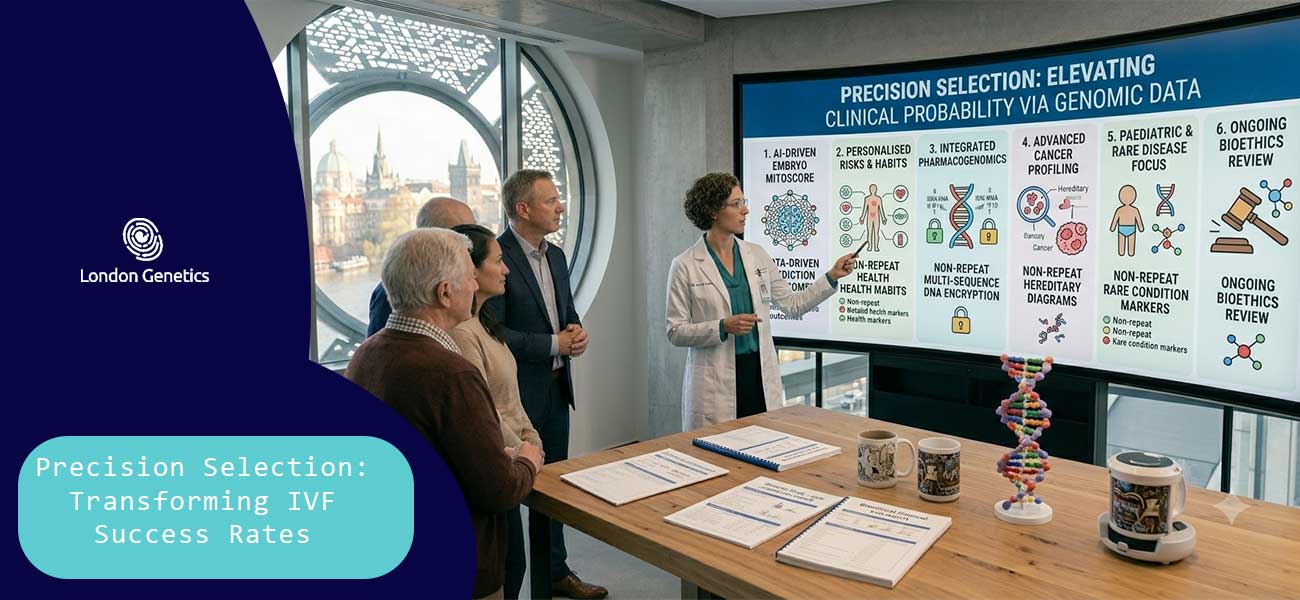
Precision Selection: Transforming IVF Success Rates
Elevating Clinical Probability via Genomic Data
In traditional fertility care, embryo selection was largely a subjective art based on fluid visual grading. Precision selection completely rewrites this clinical framework by replacing visual estimation with definitive genomic data. By ensuring that only an embryo with a perfectly balanced, 46-chromosome structure is selected, clinicians can bypass the primary biological variable that causes assisted reproductive cycles to fail, fundamentally altering the trajectory of your IVF journey.
The Direct Clinical and Statistical Impact
Integrating advanced genetic screening into the selection process yields profound improvements across every critical reproductive marker:
-
Maximising Implantation Success: An embryo with an optimal chromosomal count possesses the necessary biochemical signals to interact perfectly with the uterine lining. This precision matching drives implantation rates significantly higher compared to unscreened embryos, ensuring the blastocyst burrows securely into the endometrium.
-
Drastically Minimising Miscarriage Rates: Because the vast majority of early pregnancy losses are triggered by numerical genetic errors, transferring a verified euploid embryo directly mitigates this risk. Clinical data demonstrates that PGT-A screening drops the baseline miscarriage rate to a statistical minimum, shielding couples from the trauma of recurrent loss.
-
Reducing Time to Pregnancy: By avoiding the blind transfer of structurally compromised embryos that are destined to fail or result in early loss, couples save months of physical downtime and emotional exhaustion, achieving a healthy term pregnancy in a vastly compressed timeframe.
The Power of Single Embryo Transfer (SET)
Historically, clinicians often transferred multiple embryos simultaneously to compensate for unknown genetic viability, a practice that introduced the high medical risks of multiple pregnancies (twins or triplets). With the absolute certainty provided by precision selection, the clinical standard shifts safely to transferring a single, genetically verified embryo. This ensures the safest possible environment for both mother and child, transforming IVF from a game of chance into a highly predictable, successful medical science.
Conclusion: Embracing Scientific Certainty on Your Path Forward
Understanding that early pregnancy development is entirely dictated by a microscopic numbers game shifts the perspective on recurrent loss. Embryonic aneuploidy is not a reflection of your body’s failure to carry a child, but rather a structural cellular error that halts development naturally.
Recognising that these chromosomal imbalances are the primary driver of unexplained miscarriages allows you to move past the emotional exhaustion of unassisted cycles and look toward proven, proactive solutions.
Book Your Consultation with Dr Karimiani