Introduction to Fluorescence In Situ Hybridisation (FISH)
Fluorescence In Situ Hybridisation (FISH) stands as a cornerstone technique within the realm of molecular cytogenetics, providing researchers and clinicians with a robust method for detecting and localising the presence or absence of specific DNA sequences on chromosomes. By bridging the gap between molecular biology and traditional cytogenetics, FISH has profoundly advanced our understanding of genomic architecture and cellular pathology.
The fundamental principle of this technique relies upon the use of fluorescently labelled nucleic acid probes. These probes are meticulously synthesised to be complementary to a target genomic sequence of interest. During the procedure, both the chromosomal DNA within a cellular specimen and the probe DNA are denatured to form single strands. Subsequently, the probe is allowed to hybridise—or bind—to its specific target sequence in situ (within the intact cell or tissue architecture). Following the removal of unbound probes through rigorous washing protocols, the hybridised sequences are visualised and analysed utilising a fluorescence microscope.
Historically, traditional karyotyping techniques necessitated actively dividing cells (metaphase), which could often prove to be a laborious and time-consuming endeavour. FISH, however, offers a distinct and powerful advantage: it can be successfully performed on non-dividing (interphase) nuclei as well as metaphase chromosomes. This capability significantly expedites the diagnostic process and broadens the repertoire of biological samples that can be evaluated, including blood smears, bone marrow aspirates, and formalin-fixed, paraffin-embedded (FFPE) tissue specimens.
The clinical and investigative applications of FISH are manifold. In the field of oncology, it is routinely employed for the precise identification of chromosomal abnormalities, such as translocations, microdeletions, and gene amplifications. Beyond oncology, the technique is fundamental to Congenital Abnormalities Genetic Testing, where it facilitates the rapid detection of aneuploidies and complex structural rearrangements in both prenatal and postnatal settings. This capability has solidified its position as an essential diagnostic tool in the burgeoning field of Genomic Medicine in Paediatrics, significantly influencing patient management and genetic counselling for a new generation.
Principles and Mechanism of Fluorescence In Situ Hybridisation (FISH)
Fluorescence In Situ Hybridisation (FISH) is a powerful molecular cytogenetic technique
based on the high specificity of Watson–Crick base pairing.
It enables direct visualisation of defined DNA or RNA sequences within intact cells or chromosomes,
making it essential for detecting genomic abnormalities in clinical diagnostics and cancer genomics.
FISH begins with designing a nucleic acid probe (DNA or RNA)
that is complementary to a specific genomic region of interest.
To enable fluorescent detection, probes are labelled with fluorophores.
Labelling can be:
Direct: the dye is covalently linked to the probe itself.
Indirect: reporter molecules (e.g., biotin or digoxigenin) are incorporated
and detected later via fluorescently labelled antibodies.
Biological samples such as cells or tissue sections are fixed onto glass slides
to preserve structural integrity.
For hybridisation to occur, the double-stranded DNA in both probe and sample
must be separated.
This denaturation is achieved using optimised heat and chemical agents
(such as formamide), breaking hydrogen bonds and producing single-stranded DNA.
After denaturation, the fluorescent probe is applied to the sample.
During controlled incubation, the probe locates and binds specifically
to its complementary target sequence in situ,
within the preserved nuclear or chromosomal architecture.
This hybridisation step determines the specificity and success of the FISH assay.
Following hybridisation, the slide undergoes a series of stringent washes.
These washes remove unbound or loosely attached probes,
minimising non‑specific fluorescence.
Proper washing ensures a strong, clear signal with minimal background noise,
preventing false‑positive interpretation.
Chromosomal DNA is counterstained with dyes such as DAPI, producing a blue background
that highlights nuclear structures.
Using an epifluorescence microscope equipped with specialised filters,
fluorophores emit distinct colours (e.g., red, green, yellow) when excited.
These signals reveal the exact location and copy number of the target sequence,
allowing precise detection of genomic abnormalities.
Key Takeaway:
FISH is a highly specific, fluorescence‑based hybridisation technique
that enables direct visualisation of genomic sequences within intact cells.
Its precise detection of structural and numerical chromosomal abnormalities
makes it indispensable in cancer diagnostics, research, and personalised medicine.
Types of Probes Used in FISH Analysis
The versatility and diagnostic power of Fluorescence In Situ Hybridisation are intrinsically linked to the diverse array of probes available. The selection of a specific probe type is a critical decision, dictated entirely by the clinical or research question at hand. These molecular tools are meticulously engineered to target different genomic features, and the judicious choice among various FISH Probes is fundamental to the successful characterisation of chromosomal architecture. They can be broadly categorised into three principal classes:
1. Locus-Specific Identifier (LSI) Probes
Locus-specific probes are designed to hybridise to a unique, specific sequence of DNA, such as a single gene or a defined chromosomal region. They are amongst the most frequently used probes in both diagnostic and research settings. Their applications are extensive and include:
- Detection of Microdeletions/Microduplications: Identifying the gain or loss of small segments of DNA that are too diminutive to be resolved by conventional cytogenetics.
- Gene Amplification Analysis: Quantifying the copy number of specific oncogenes, a classic example being the assessment of HER2 gene amplification in breast cancer specimens to guide therapeutic decisions.
- Translocation and Gene Fusion Detection: Utilising strategies such as “dual-colour fusion” or “break-apart” probes to pinpoint chromosomal rearrangements. The detection of the BCR-ABL1 gene fusion in Chronic Myeloid Leukaemia (CML) is a prime example of their diagnostic utility.
2. Centromeric Probes (CEP)
Also known as Chromosome Enumeration Probes, CEP probes are designed to bind to the highly repetitive alpha-satellite DNA sequences located at the centromere of a specific chromosome. As each chromosome possesses a unique centromeric sequence, these probes provide a rapid and highly reliable method for chromosome counting (enumeration) within interphase nuclei. Consequently, their primary application is the diagnosis of aneuploidy—an abnormal number of chromosomes. They are routinely employed in prenatal and postnatal testing for conditions such as Trisomy 21 (Down syndrome), Trisomy 18 (Edwards syndrome), and aneuploidies of the sex chromosomes.
3. Whole Chromosome Painting (WCP) Probes
Whole Chromosome Painting probes consist of a complex cocktail or library of smaller probes that, collectively, bind along the entire length of a particular chromosome. This effectively “paints” the target chromosome with a single, uniform fluorescent colour. WCP probes are invaluable for identifying complex and subtle structural rearrangements, such as cryptic translocations between different chromosomes. They are also instrumental in characterising marker chromosomes—small, unidentified fragments of chromosomal material—by unequivocally determining their chromosomal origin. This makes them a powerful tool for analysing complex karyotypes often observed in tumours and congenital disorders.
Methodology and Laboratory Procedure of FISH
The successful execution of Fluorescence In Situ Hybridisation (FISH) relies on a carefully standardised laboratory workflow.
Although the conceptual principles are straightforward, practical implementation requires precise control of experimental conditions.
Each stage of the procedure must be optimised to produce strong, specific fluorescent signals while minimising background noise.

Explore the main laboratory stages of the FISH procedure.

This first stage prepares cells or tissue sections for probe binding.
Procedures vary depending on the sample type such as blood lymphocytes,
amniocytes, or formalin‑fixed paraffin‑embedded (FFPE) tissues.
FFPE samples are first deparaffinised with xylene
and rehydrated through graded ethanol washes.
Permeabilisation is then performed using enzymatic digestion
(e.g., proteinase K or pepsin) to remove proteins
and allow access to chromosomal DNA.
Finally, the sample is fixed again,
commonly using formaldehyde,
to preserve nuclear and chromosomal structure.

For hybridisation to occur,
both the probe DNA and the target chromosomal DNA
must be converted into single strands.
This is typically achieved by incubating the slide
in a denaturing solution containing formamide
at elevated temperatures around 70–75°C.
The probe mixture is denatured separately
before being applied to the prepared sample.

The denatured probe solution is applied to the slide
and the area is sealed to prevent evaporation.
The slide is then incubated in a humidified chamber,
typically at 37°C,
for several hours or overnight.
During this time the probe
locates and anneals specifically
to its complementary DNA sequence
within the chromosome or nucleus.

Following incubation,
the slide is washed to remove any unbound
or non‑specifically attached probe molecules.
These washes are performed under carefully controlled conditions
using buffers such as saline‑sodium citrate (SSC).
Higher wash stringency
ensures that only perfectly matched probe‑target hybrids remain,
producing clear and reliable fluorescence signals.

In the final step,
the specimen is counterstained with a DNA dye,
most commonly DAPI,
which fluoresces blue and highlights all cell nuclei.
An anti‑fade mounting medium is added
to preserve fluorescence signals.
The slide is then examined using a fluorescence microscope
equipped with appropriate optical filters
to detect the coloured signals emitted by the probes.
Key Takeaway:
The FISH laboratory workflow consists of carefully controlled stages,
including sample preparation, denaturation, hybridisation,
stringent washing, and fluorescent visualisation.
Accurate optimisation of each step is essential
to obtain clear signals and reliably detect genomic abnormalities.
Clinical and Research Applications of FISH
The versatility of Fluorescence In Situ Hybridisation has rendered it an indispensable tool, bridging the gap between fundamental research and routine clinical diagnostics. Its ability to provide rapid, specific, and visually compelling information about genomic architecture has cemented its role in numerous fields. The applications of FISH are extensive, spanning from the detection of constitutional abnormalities to the molecular characterisation of complex cancers.
1. Oncology and Haematology:
In clinical oncology, FISH is pivotal for diagnosis, prognostication, and guiding targeted therapies.
- Solid Tumours: It is routinely used to detect gene amplifications, such as the assessment of HER2 gene status in breast and gastric cancers, which directly informs the decision to use trastuzumab therapy.
- Haematological Malignancies: The technique is the gold standard for identifying hallmark chromosomal translocations. The detection of the t(9;22) translocation, forming the BCR-ABL1 fusion gene in Chronic Myeloid Leukaemia (CML), is a classic example of the diagnostic power of FISH. Similarly, it is crucial for identifying rearrangements involving genes like PML-RARA in acute promyelocytic leukaemia.
2. Prenatal and Postnatal Genetic Diagnosis:
FISH has revolutionised cytogenetics by offering a rapid method for detecting aneuploidy (abnormal chromosome numbers) in uncultured cells.
- Rapid Aneuploidy Screening: Using probes for chromosomes 13, 18, 21, X, and Y on amniocytes or chorionic villus samples allows for the swift prenatal detection of conditions like Down syndrome (Trisomy 21), Edwards syndrome (Trisomy 18), and Turner syndrome (Monosomy X).
- Microdeletion Syndromes: It is the primary diagnostic tool for syndromes caused by the loss of tiny chromosomal segments, such as the 22q11.2 deletion in DiGeorge syndrome, which are undetectable by standard karyotyping.
3. Basic and Translational Research:
Beyond the clinic, the application of FISH in research has yielded profound insights into genome biology.
- Gene Mapping: It has been instrumental in physically localising newly discovered genes to specific chromosomal bands.
- Genome Organisation: Researchers use multi-colour FISH (M-FISH) and spectral karyotyping (SKY) to study complex chromosomal rearrangements and the spatial organisation of chromosomes within the interphase nucleus (chromosome territories).
- Comparative Genomics: By applying probes from one species to the chromosomes of another (zoo-FISH), scientists can study evolutionary relationships and chromosomal rearrangements over time.
In summary, from confirming a clinical diagnosis that directly impacts patient care to mapping the very structure of our genome, the broad utility of the technique is undeniable. Consequently, the continued refinement of the FISH methodology ensures its enduring relevance in the era of genomic medicine.
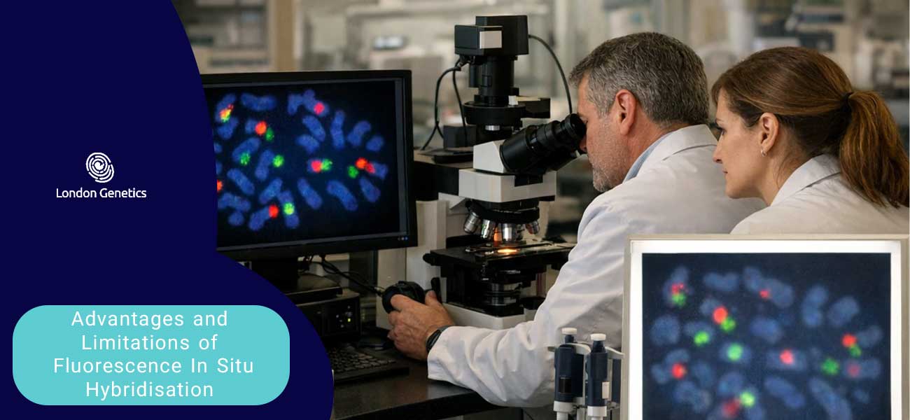
Advantages and Limitations of Fluorescence In Situ Hybridisation
While Fluorescence In Situ Hybridisation has revolutionised molecular cytogenetics, it is essential for researchers and clinicians to possess a balanced understanding of its capabilities and constraints. Like any powerful scientific technique, its utility is defined by both its profound advantages and its inherent limitations. A careful evaluation of these factors is crucial for its appropriate application.
Advantages
- Rapidity and Interphase Analysis: One of the most significant advantages over traditional karyotyping is the ability to perform analysis on non-dividing (interphase) nuclei. This circumvents the need for cell culture, a process that can be time-consuming and sometimes unsuccessful. Consequently, results can often be obtained within 24-48 hours, a critical factor in time-sensitive clinical situations such as prenatal diagnosis.
- High Specificity and Sensitivity: The technique is exceptionally specific, as probes are designed to bind only to their exact complementary DNA sequence. This allows for the unambiguous detection of specific genes or chromosomal regions with a very low probability of false positives when performed correctly.
- Versatility of Sample Types: FISH can be successfully applied to a diverse range of specimen types, including fresh or cultured cells, blood and bone marrow smears, and, importantly, formalin-fixed, paraffin-embedded (FFPE) tissue sections. This latter capability makes it an invaluable tool for retrospective studies on archived pathological material.
- Preservation of Morphological Context: As the analysis is performed in situ, the genetic information is viewed within the context of the intact cell and tissue architecture. This allows for the correlation of genetic abnormalities with specific cell types, providing a layer of spatial information that is lost in extraction-based molecular methods like PCR.
Limitations
- Targeted, Not a Discovery Tool: A fundamental limitation of FISH is its targeted nature. It can only detect the abnormalities for which a specific probe is used. One must have prior knowledge of the chromosomal region of interest. Consequently, it cannot be used to identify novel or unexpected genomic rearrangements across the entire genome in the way that techniques like whole-genome sequencing can.
- Limited Genomic Resolution: While excellent for detecting copy number changes and rearrangements, the technique cannot resolve alterations at the DNA sequence level. It is incapable of detecting point mutations, small insertions or deletions, or epigenetic modifications.
- Technical and Cost Considerations: The procedure requires specialised and expensive equipment, notably a high-quality fluorescence microscope with appropriate filter sets. The probes themselves can be costly, and the interpretation of results requires significant expertise to avoid misdiagnosis due to technical artefacts, such as weak signals or background noise.
Ultimately, the decision to employ Fluorescence In Situ Hybridisation must be made by carefully weighing its profound diagnostic advantages against its inherent limitations in the context of the specific scientific or clinical question being addressed.
Conclusion: The Enduring Legacy and Future of FISH
In conclusion, the legacy of Fluorescence In Situ Hybridisation is secure.
Its fundamental strength—the ability to visualise the genome directly
within its cellular and morphological context—remains a unique and powerful attribute
that extraction-based methods like next-generation sequencing cannot replicate.
The ongoing innovations in automation, probe chemistry, and multiplexing
are not merely incremental improvements;
they represent a fundamental evolution of the technique,
ensuring it keeps pace with the demands of modern molecular biology.
As we advance further into the era of precision medicine and systems biology,
a more powerful, quantitative, and spatially resolved
Fluorescence In Situ Hybridisation will undoubtedly remain an indispensable tool,
illuminating the complexities of the genome
and guiding clinical decisions with ever-increasing clarity and precision.
Looking ahead, continued innovation will strengthen the technique in key areas:
- Enhanced automation and quantitative analysis
- Improvements in probe chemistry and signal amplification
- Higher multiplexing for spatial genomics applications
- Integration with super-resolution microscopy
- Greater utility in precision medicine and diagnostics
Why This Matters:
FISH provides unique visual context to genomic data that other methods lack.
This ability to see genetic information within the cell is critical
for accurate diagnosis, understanding disease mechanisms,
and developing targeted therapies in modern medicine.
The Value of Continued Innovation:
Ongoing advancements are transforming FISH from a targeted diagnostic tool
into a powerful platform for large-scale discovery.
This evolution ensures its central role in answering complex questions
in molecular biology and clinical genetics for years to come.
Your Next Step:
Discover how advanced FISH methodologies are being integrated
into cutting-edge clinical diagnostics,
pharmaceutical research,
and the field of spatial biology.