
Introduction: Understanding Neural Tube Defects (NTDs)
Embarking on the journey of pregnancy brings a wave of excitement, but it is completely natural for expectant parents to feel a sense of anxiety about their baby’s health. One of the key areas focused on during early pregnancy care is the prevention and detection of neural tube defects (NTDs). These are serious structural conditions that affect the development of a baby’s brain and spine during the first few weeks of pregnancy.
While hearing terms like spina bifida can feel overwhelming, understanding the underlying causes and knowing your options can significantly reduce your anxiety. This is where prenatal genetic counselling plays a vital role. By combining advanced medical insights with compassionate guidance, genetic professionals help families navigate risks, understand screening options, and make informed decisions for a healthier future. In this guide, we will break down what causes these defects, how they are detected, and the proactive steps you can take to protect your growing family.
What Causes Neural Tube Defects? (The Primary Triggers)
The neural tube normally folds and closes completely by the 28th day after conception. When this structure fails to close properly, neural tube defects (NTDs) occur. Medical research shows these defects are multifactorial, triggered by a complex combination of genetic predispositions and environmental influences.
Neural tube defects sit at the intersection of genetic blueprints and environmental biochemistry. Recognizing risk factors like MTHFR mutations or folate deficits allows families to use individual genetic counselling to map out proactive, highly effective preconception prevention strategies.
Common Types of NTDs: Spina Bifida and Beyond
Not all neural tube defects affect the body in the same way. Depending on where the neural tube fails to close along the embryonic spine or head, the resulting condition can range from mild physical challenges to severe, life-threatening complications.
By understanding the distinct types of NTDs, families can better grasp what certain ultrasound findings mean and what kind of specialized care or interventions might be required.
Spina Bifida: What You Need to Know
Spina bifida—which literally means “split spine”—is the most common type of neural tube defect. It occurs when the lower part of the neural tube fails to close completely, leaving the spinal cord and backbones poorly formed. Spina bifida generally presents in three main forms:
-
Spina Bifida Occulta: Often called “hidden” spina bifida, this is the mildest and most common form. There is a small gap in the spinal bones, but the spinal cord and nerves remain completely intact inside. Most people with this form never even know they have it, as it rarely causes disabilities or visible symptoms.
-
Meningocele: In this rarer form, a fluid-filled sac pushes out through the opening in the baby’s back. Fortunately, the spinal cord itself is not inside this sac, meaning nerve damage is usually minimal, and it can often be repaired with surgery after birth.
-
Myelomeningocele: This is the most severe and clinically significant form of spina bifida. Here, the spinal cord and nerve pathways actually push through the open spinal column into the sac on the baby’s back. This exposure causes moderate to severe nerve damage, which can lead to complications such as partial or total paralysis of the legs, bladder and bowel dysfunction, and hydrocephalus (fluid buildup in the brain).
Anencephaly and Other Severe Forms
While spina bifida primarily impacts the spine, other types of NTDs occur at the upper end of the neural tube, affecting the development of the skull and brain:
-
Anencephaly: This is a severe and heartbreaking form of NTD that occurs when the upper portion of the neural tube fails to close. As a result, the baby’s brain, skull, and scalp do not develop completely. Infants born with anencephaly are missing major parts of the forebrain and cerebrum, the areas responsible for thinking and coordinating movement. Sadly, this condition is fatal, and most babies survive for only a few hours or days after birth.
-
Encephalocele: This is a much rarer type of defect where a sac-like protrusion of the brain and the membranes covering it breaks through an opening anywhere in the skull (most commonly at the back of the head). The severity of encephalocele depends heavily on its size, location, and how much brain tissue is pushed outside the skull.
The Power of Prevention: The Crucial Role of Folic Acid
While you cannot alter your genetic makeup, ensuring an adequate intake of folic acid is a highly effective, scientifically proven step to dramatically lower the risk of neural tube defects (NTDs), preventing up to 70 per cent of all clinical cases.

Maintaining a folate-rich diet and establishing a proper supplementation routine before conception safeguards embryonic growth during the critical early windows of development.


Key Takeaway:
Folic acid represents a cornerstone of modern preconception care. By matching daily supplementation—whether standard 400mcg or therapeutic 5mg—to individual genetic baselines and MTHFR variants, families can actively intervene to maximize embryonic protection.
How Prenatal Genetic Counselling Helps Expectant Parents
While public health guidelines emphasize preventive steps like taking folic acid, navigating individual risks for congenital conditions can still feel incredibly complex. This is exactly where professional genetic guidance steps in. A genetic counsellor is a specialized healthcare professional who translates intricate genetic data into clear, actionable, and compassionate steps to help families assess their risk of neural tube defects.
Rather than offering a one-size-fits-all approach, a genetic consultation provides a deeply personalized roadmap. It helps families move away from generalized statistics and focus on their specific biological realities regarding neural tube defects.
Evaluating Family History and Neural Tube Defects
One of the most powerful tools a genetic counsellor uses to evaluate the risk of neural tube defects is the construction of a detailed medical family tree spanning at least three generations. This systematic process is far more thorough than a standard medical history questionnaire:
-
Identifying Hereditary Patterns: The counsellor maps out health conditions, spinal issues, and previous occurrences of neural tube defects on both the maternal and paternal sides to see if a genetic pattern exists.
-
Evaluating Consanguinity Risks: In families where marriages between relatives are common, the counsellor calculates the likelihood of inheriting recessive genes that could potentially contribute to the development of neural tube defects.
-
Pinpointing High-Risk Recurrence: If a couple has already experienced a pregnancy affected by an NTD, the counsellor evaluates the specific circumstances to determine the statistical likelihood of neural tube defects happening again in subsequent pregnancies.
Beyond the Chart: What Does a Genetic Specialist Do?
Constructing a family tree is just the beginning. A specialized session focused on neural tube defects involves several critical steps designed to empower and support parents:
-
Calculating Personalized Risk: Instead of looking at generic internet statistics, the expert combines your family history, maternal age, lifestyle factors, and genetic markers to calculate a precise risk percentage for neural tube defects in your specific pregnancy.
-
Coordinating Advanced Testing: If an elevated risk for neural tube defects is identified, the specialist serves as an expert guide, determining exactly which advanced screening tools or diagnostic options are appropriate for your situation.
-
Providing Objective, Compassionate Support: Specialists explain the complex pros, cons, and accuracy rates of tests used to detect neural tube defects, helping you make informed, autonomous choices that align with your family’s personal values and ethical beliefs.
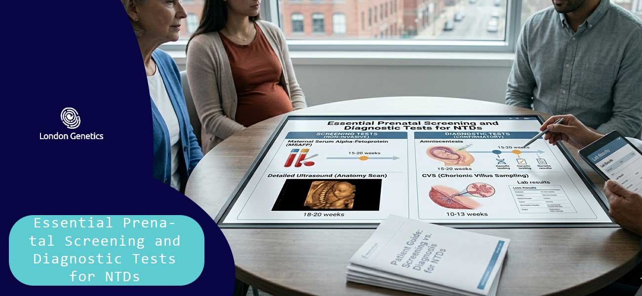
Essential Prenatal Screening and Diagnostic Tests for NTDs
When assessing the risk of neural tube defects, modern medicine provides highly accurate tools to evaluate the development of the fetal spine and brain. These clinical evaluations are divided into two main categories: screening tests (which calculate the statistical probability of a condition) and diagnostic tests (which provide a definitive yes-or-no answer).
By utilizing these specialized medical technologies, healthcare providers can closely monitor a pregnancy and detect the presence of neural tube defects early in gestation.
Maternal Serum Alpha-Fetoprotein (MSAFP) Screening
The MSAFP test is a simple blood test offered to pregnant individuals, typically between the 15th and 20th weeks of pregnancy, and serves as a primary tool for flag-raising:
-
Understanding Alpha-Fetoprotein: AFP is a protein naturally produced by the yolk sac and fetal liver. A small, predictable amount of this protein normally crosses the placenta into the mother’s bloodstream.
-
The Link to Neural Tube Defects: If the fetal spinal column or skull has an open abnormality, extra amounts of AFP leak into the amniotic fluid and enter the maternal blood. Consequently, significantly elevated levels of MSAFP often indicate an increased risk of open neural tube defects, such as spina bifida or anencephaly.
-
A Tool for Probability: It is important to note that an abnormal MSAFP result is a screening indicator, not a definitive diagnosis of neural tube defects. It simply signals the need for further, more detailed testing.
Advanced High-Resolution Ultrasound
If a blood screening shows an elevated risk, or if there is a known family history of developmental complications, a targeted, high-resolution ultrasound (often called an anomaly scan) is performed between the 18th and 22nd weeks:
-
Visualizing the Fetal Anatomy: Using advanced imaging technology, a maternal-fetal medicine specialist performs a detailed head-to-toe examination, focusing heavily on the structural integrity of the fetal cranium and spinal column to visually rule out neural tube defects.
-
Detecting Brain and Spine Markers: High-resolution ultrasound can clearly identify physical markers of neural tube defects. This includes visualizing open lesions along the vertebrae or detecting specific changes in brain shape (often referred to as the “lemon” or “banana” signs) caused by spinal fluid shifts.
-
High Diagnostic Accuracy: In expert hands, modern ultrasound imaging can detect over 95% of open neural tube defects, making it an indispensable, non-invasive tool for expectant parents.
Diagnostic Tests: Amniocentesis
When screening results are ambiguous or indicate a high probability of structural complications, a definitive diagnostic procedure called amniocentesis may be recommended:
-
Analyzing Amniotic Fluid: During this procedure, a specialist uses a thin needle under ultrasound guidance to extract a small sample of fluid from the amniotic sac surrounding the baby.
-
Testing for AFAFP and AChE: The extracted fluid is tested directly for elevated levels of Amniotic Fluid Alpha-Fetoprotein (AFAFP) and Acetylcholinesterase (AChE). The presence of AChE in the fluid is a highly specific chemical signature that confirms an open neural tube tissue exposure.
-
Confirming Neural Tube Defects: Unlike blood screenings, the biochemical results of an amniocentesis provide a highly definitive diagnosis, allowing healthcare providers to confirm or rule out neural tube defects with near-absolute certainty.
Conclusion: Empowering Your Pregnancy Journey with Knowledge
Navigating prenatal health choices can feel overwhelming, but building a foundational understanding of neural tube defects (NTDs) is a powerful step toward a proactive and confident pregnancy. While embryonic structural complexities are serious, modern medicine provides remarkable pathways for early prevention, accurate screening, and structured management.
You do not have to carry the weight of reproductive uncertainty alone. Advanced prenatal screening and expert genetic guidance exist to offer absolute clarity, definitive answers, and reduced anxiety for expectant parents, allowing you to focus on the joy of welcoming a new life.
Proactive Journey:
Transforming maternal healthcare from a position of anxiety to one of absolute confidence through structured, evidence-based prenatal knowledge.
Clinical Prevention:
Utilising well-timed, highly precise folic acid supplementation routines to actively block the primary environmental triggers of neural tube defects.
Advanced Screenings:
Leveraging high-resolution anomaly scans and targeted testing options to detect embryonic development patterns with extreme clinical accuracy.
Empathetic Guidance:
Relying on specialized prenatal consultations to dismantle the stress of genetic uncertainties and secure a clear, customized roadmap.
Informed Family Planning:
Taking calculated, practical preconception steps today to safeguard fetal health and offer the maximum biological protection for your future child.