What Is Trophectoderm Biopsy in IVF?
A trophectoderm biopsy is a specialised procedure performed during the later stages of IVF to assess the genetic health of an embryo before implantation. At the blastocyst stage—typically day five or six of development—the embryo naturally forms two distinct cell groups: the inner cell mass (which becomes the baby) and the trophectoderm (which forms the placenta). During a Trophectoderm Biopsy, a small number of trophectoderm cells are gently removed for advanced genetic analysis.
Because these cells are destined to form the placenta rather than the fetus, this technique offers a highly informative and safer method of evaluating embryo quality.
This approach sits at the forefront of Reproductive Genetics, allowing clinicians to thoroughly analyse embryos without disrupting their developmental potential.
A trophectoderm biopsy is most commonly used alongside Preimplantation Genetic Testing, providing detailed insight into chromosomal and genetic conditions. By identifying the healthiest embryos, fertility specialists can improve implantation rates, reduce the likelihood of miscarriage, and support safer, more successful IVF outcomes.
Overall, a Trophectoderm Biopsy is a precise, modern, and reliable tool that enhances embryo selection while maintaining the integrity of embryonic development.
Why Trophectoderm Biopsy Is Performed During IVF
Trophectoderm Biopsy is an advanced laboratory procedure used during IVF to assess the genetic health of embryos before transfer. By analysing a small sample of cells from the outer layer of the embryo, specialists can identify chromosomal abnormalities and select embryos with the best potential for a healthy pregnancy.
By enabling detailed genetic screening before embryo transfer, Trophectoderm Biopsy reduces uncertainty in embryo selection and plays a key role in modern IVF strategies aimed at achieving healthy pregnancies.
How the Trophectoderm Biopsy Procedure Works
The Trophectoderm Biopsy procedure is a carefully controlled laboratory technique performed during the IVF process to obtain a small sample of cells from a developing embryo. This procedure is typically carried out when the embryo reaches the blastocyst stage, around day five or six after fertilisation, when the embryo has developed distinct cellular structures.
At this stage, the embryo contains two main cell groups: the inner cell mass, which will develop into the fetus, and the trophectoderm layer, which later forms the placenta. During a Trophectoderm Biopsy, embryologists remove a few cells from the trophectoderm layer while leaving the inner cell mass untouched.
The procedure generally follows several key steps:
-
Blastocyst Development
After fertilisation, embryos are cultured in the laboratory for several days until they reach the blastocyst stage, when the trophectoderm cells become accessible for biopsy.
-
Creating a Small Opening in the Embryo Shell
A highly precise laser is used to create a tiny opening in the zona pellucida, the protective outer layer surrounding the embryo.
-
Removing Trophectoderm Cells
Using a specialised microscopic pipette, the embryologist gently extracts a small group of trophectoderm cells, usually between five and ten cells. This step constitutes the actual Trophectoderm Biopsy.
-
Genetic Analysis
The collected cells are then sent for genetic evaluation, often through Preimplantation Genetic Testing, to detect chromosomal abnormalities or inherited genetic conditions.
-
Embryo Cryopreservation
In many cases, the embryo is frozen using vitrification while the genetic results are being analysed. Once results are available, the healthiest embryo can be selected for transfer in a later IVF cycle.
A Trophectoderm Biopsy requires a high level of precision and advanced laboratory expertise. When performed by experienced embryologists, the procedure is considered safe and has minimal impact on embryo development. This technique allows fertility specialists to gain valuable genetic information that can significantly improve embryo selection and IVF success rates.
The Role of Trophectoderm Biopsy in Preimplantation Genetic Testing (PGT)
Trophectoderm Biopsy provides the essential cell sample used in Preimplantation Genetic Testing (PGT), allowing specialists to examine an embryo’s chromosomal and genetic profile before transfer. Because the sampled cells come from the outer layer of the blastocyst, the procedure provides reliable genetic information without disturbing the fetal‑forming inner cell mass.

How Trophectoderm Biopsy supports different types of PGT.


Key Takeaway:
Trophectoderm Biopsy is a foundational tool in modern PGT, providing the high‑quality genetic information needed to guide embryo selection and enhance the safety and success of IVF treatments.
Benefits of Trophectoderm Biopsy for Embryo Selection
Trophectoderm Biopsy offers several important advantages in the embryo selection process during IVF. By allowing specialists to analyse the genetic makeup of embryos before transfer, this technique helps identify those with the highest potential for healthy development and successful implantation.
One of the main benefits of Trophectoderm Biopsy is the ability to improve the accuracy of embryo selection. Traditional embryo assessment mainly relies on visual evaluation of embryo shape, cell division, and growth patterns under the microscope. While these indicators provide useful information, they cannot reveal underlying chromosomal abnormalities. By obtaining a small sample of cells from the trophectoderm layer, genetic testing can provide deeper insight into the embryo’s genetic health.
Another key advantage is the reduction of chromosomal abnormalities in transferred embryos. Through Preimplantation Genetic Testing (PGT) performed on cells collected during a Trophectoderm Biopsy, clinicians can detect embryos with the correct number of chromosomes. Selecting chromosomally normal embryos may increase implantation rates and reduce the likelihood of miscarriage.
Trophectoderm Biopsy can also support single embryo transfer strategies. When doctors have reliable genetic information about embryo viability, they can confidently transfer one high‑quality embryo rather than multiple embryos. This approach helps lower the risk of multiple pregnancies, which are associated with higher medical risks for both the mother and the babies.
In addition, this technique can be particularly beneficial for certain groups of patients, including women of advanced maternal age, couples with a history of repeated IVF failure, and those with recurrent pregnancy loss. In these situations, Trophectoderm Biopsy can provide valuable genetic data that helps guide treatment decisions and improve clinical outcomes.
Overall, the use of Trophectoderm Biopsy in embryo selection allows fertility specialists to make more informed choices, increasing the likelihood of a successful pregnancy while supporting safer and more personalised IVF treatment.
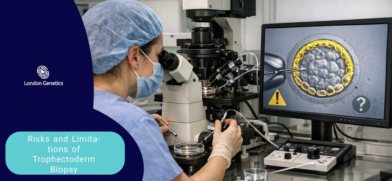
Risks and Limitations of Trophectoderm Biopsy
Although Trophectoderm Biopsy is widely used in modern IVF laboratories and is generally considered safe, it is important to recognise that the procedure has certain risks and limitations. Understanding these factors helps patients and clinicians make informed decisions when considering genetic testing during IVF.
One potential risk is the possibility of minor damage to the embryo during the biopsy process. The procedure involves removing a small number of cells from the trophectoderm layer using specialised microscopic tools and a laser. While experienced embryologists perform this technique with great precision, there is still a very small chance that the embryo’s development could be affected.
Another limitation relates to embryo mosaicism. In some embryos, not all cells have the same genetic makeup. Because a Trophectoderm Biopsy samples only a small group of cells from the outer layer of the embryo, the genetic results may not always perfectly represent the entire embryo. In rare cases, this could lead to uncertain or inconclusive test results.
There is also the possibility that the biopsy may not yield enough high‑quality DNA for accurate genetic testing. If the collected cells do not provide sufficient genetic material, additional analysis may be required, or the embryo may not receive a clear genetic diagnosis.
In addition, the process typically requires embryo freezing. After a Trophectoderm Biopsy, embryos are often cryopreserved while genetic testing is performed. Although modern freezing techniques such as vitrification are highly effective, this additional step can extend the IVF timeline and may not be suitable for every patient.
Finally, it is important to note that Trophectoderm Biopsy and subsequent genetic testing cannot guarantee a successful pregnancy or the birth of a completely healthy baby. While the procedure can detect many chromosomal abnormalities and certain genetic conditions, it does not screen for all possible health issues.
For these reasons, Trophectoderm Biopsy should be considered as one component of a comprehensive fertility treatment plan, combined with professional medical guidance and careful clinical evaluation.
Trophectoderm Biopsy in IVF: Insights, Benefits, and the Future of Genetic Screening
Trophectoderm Biopsy has become a cornerstone of modern reproductive medicine, enabling specialists to analyse embryo genetics before transfer. When paired with Preimplantation Genetic Testing (PGT), it helps identify embryos with the greatest potential for healthy development, successful implantation, and reduced risks during pregnancy.
As accuracy improves and laboratory techniques advance, Trophectoderm Biopsy continues to enhance personalised fertility care—supporting informed decisions and more confident IVF journeys.
Informed Embryo Selection:
Genetic analysis provides clear insight into chromosomal health and inherited conditions, helping clinicians choose embryos with the highest chance of success.
Improved IVF Success Rates:
By detecting abnormalities early, Trophectoderm Biopsy supports safer single‑embryo transfer, lowers miscarriage risk, and increases implantation potential.
Understanding Limitations:
Factors such as mosaicism, the need for freezing, and occasional inconclusive results highlight the value of expert counselling and realistic expectations.
Empowering Patients Through Knowledge:
Clear explanations of benefits and limitations help individuals and couples make informed decisions about whether PGT and biopsy are right for them.
A Growing Role in Reproductive Genetics:
With continuous advancements in technology, Trophectoderm Biopsy is expected to become even more precise—refining embryo selection and shaping the future of IVF success.