Introduction: The Architecture of Acquired Genetic Changes
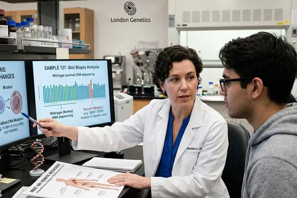
Every single day, the human body orchestrates billions of complex cellular divisions, replicating vast amounts of genetic code with astonishing fidelity. Yet, despite the presence of highly sophisticated cellular repair mechanisms, this biological duplication process is not completely flawless. Over the course of a lifetime, subtle alterations can spontaneously manifest within the DNA of individual non-reproductive cells. These changes, scientifically categorized as somatic mutations, act as quiet, localized shifts within the body’s internal architecture. Unlike the genetic traits we are born with, these variations are entirely acquired, developing progressively as a consequence of cellular aging or prolonged environmental exposure.
Understanding the true nature of somatic mutations requires a fundamental shift in how we perceive genetic anomalies. Historically, many people associated genetic disease exclusively with inheritance—the passing of faulty traits from parent to offspring. However, modern molecular biology has revealed that the vast majority of cellular errors are entirely non-inherited. Because somatic mutations are confined strictly to specific body tissues, they do not impact the wider germline, meaning they can never be transmitted to future generations. They remain isolated events, locked within the specific cellular lineage where the initial copying error occurred.
┌────────────────────────────────────────────────────────────────────────┐
│                   THE PATHWAY OF ACQUIRED CELLULAR ERRORS              │
├────────────────────────────────────────────────────────────────────────┤
│  [Environmental/Aging Triggers] ──> Random DNA Copying Error           │
│                                              │                         │
│                                              ▼                         │
│  [Localized Tissue Proliferation] ─> Somatic Mutations Formed          │
│                                              │                         │
│                                              ▼                         │
│  [Clinical Precision Analysis] ────> Somatic (Tumour) Genomic Testing   │
└────────────────────────────────────────────────────────────────────────┘
This hidden, localized behaviour poses a unique challenge for modern diagnostic medicine. Because these acquired variations are absent from a patient’s healthy blood or saliva, traditional hereditary screening panels cannot track them down. To truly capture these elusive variants and understand how they alter tissue behaviour, specialists must look directly at the affected site through advanced somatic (tumour) genomic testing. By evaluating the precise molecular footprint left by these somatic mutations, healthcare providers can move past theoretical probabilities and uncover the exact, real-time genetic drivers governing a patient’s current health profile.
What Are Somatic Mutations? (The Cellular Blueprint)
Somatic mutations are acquired genetic alterations that occur in the DNA of non-reproductive cells after conception. Unlike inherited variants, these changes develop during a person’s lifetime, remain confined to specific tissues, and cannot be passed down to offspring, typically arising from spontaneous replication errors or environmental exposures.
Recognising that somatic mutations accumulate silently across a lifetime underscores the importance of analysing the cell’s internal copying mechanics. By understanding how typographical slips bypass proofreading systems during mitosis, clinical research can better map how these localized genomic shifts alter cell behaviour and drive tissue transformation.
Somatic vs. Germline Variants: Mapping the Critical Differences
In the field of clinical genomics, drawing a definitive line between different types of genetic variations is paramount to understanding disease progression. Patients frequently look at their laboratory reports and struggle to comprehend the practical differences between an inherited defect and an acquired one. To establish absolute clarity, clinicians break down the human genetic landscape into two distinct cellular domains: the somatic track and the germline track. Conducting a comprehensive somatic vs germline analysis is not just an academic exercise; it is a vital diagnostic necessity that dictates how a patient’s condition is managed, how their family members are screened, and how their therapeutic roadmap is constructed.
The fundamental divergence between these two groups of genetic variants comes down to the origin of the cell, its location within the body, and its evolutionary destiny.
                  ┌────────────────────────────────────────┐
                  │      THE GENETIC ORIGIN DIVERGENCE     │
                  └───────────────────┬────────────────────┘
                                      │
         ┌────────────────────────────┴────────────────────────────┐
         ▼                                                         ▼
┌─────────────────────────────────┐               ┌─────────────────────────────────┐
│       GERMLINE MUTATIONS        │               │        SOMATIC MUTATIONS        │
│ • Inherited via sperm/egg       │  VS (Origins) │ • Acquired post-conception     │
│ • Present in EVERY single cell   │               │ • Present ONLY in local tissue  │
│ • Transmissible to offspring    │               │ • Cannot be passed to children  │
└─────────────────────────────────┘               └─────────────────────────────────┘
The Inherited Blueprint: Germline Mutations
To understand the contrast, we must first look at hereditary genetics. Germline mutations originate directly within the reproductive cells—the sperm or the egg—of a parent. Because these errors are present at the absolute moment of conception, they are hardcoded into the initial zygote. As that single cell divides and develops into a complete human being, the initial error is faithfully copied into every single tissue.
Consequently, if an individual carries germline mutations (such as a pathogenic BRCA1 or Lynch Syndrome variant), that abnormality can be found in their blood, saliva, skin, and organs. Because these variants reside within their own reproductive system, there is a permanent 50% chance of passing this inherited vulnerability down to their children, making family-wide screening and genetic counselling absolutely crucial.
The Acquired Event: Somatic Mutations
In stark contrast, somatic mutations develop much later in life and are completely independent of parental heritage. These variations occur in non-reproductive cells long after birth, often triggered by localized cellular fatigue or external carcinogens. A patient is not born with these changes; instead, a single cell in a specific organ undergoes a spontaneous genetic shift.
Because this event is isolated, the resulting somatic mutations are confined strictly to that specific cell and its direct anatomical descendants. If a woman develops an acquired mutation in a single breast tissue cell, that alteration remains locked within that localized cluster. Her healthy blood cells, her lung tissue, and—most importantly—her egg cells remain completely untouched by this error. Therefore, these variants are entirely non-transmissible; they cannot be passed down to offspring, meaning they pose zero genetic risk to the patient’s children or siblings.
Clinical Implications of Somatic vs Germline Testing
Differentiating between these two pathways alters the entire clinical strategy. When an oncologist runs a test and identifies germline mutations, the focus extends beyond the patient to protect the entire family tree, as siblings and children may carry the same silent threat. Conversely, identifying isolated somatic mutations shifts the medical focus entirely to the individual’s localized condition. It tells the medical team that the issue is an isolated, acquired phenomenon, requiring targeted intervention directly at the site of the altered tissue without the need for multi-generational family screening.
Environmental Triggers and Cellular Errors: What Causes These Mutations?
The genesis of acquired genetic variants is rarely a single, isolated event. Instead, the development of somatic mutations is driven by a complex interplay between internal biological vulnerabilities and external exposures. While our bodies possess remarkable defensive mechanisms, the continuous accumulation of these structural DNA alterations over a lifetime remains practically inevitable.

Understanding the root causes of genetic mutations requires evaluating both the internal replication errors made during cell maintenance and the external environmental pressures that accelerate this decay.


Key Takeaway:
Analysing the hard science behind somatic variations reveals that your genetic stability is shaped by both time and environment. Mapping how intrinsic replication inefficiencies interact with lifestyle mutagens allows clinical teams to move past general assumptions and design targeted, protective wellness programmes based on your real cellular exposure history.
The Oncological Connection: How Acquired DNA Changes Drive Tumour Growth
To appreciate the true clinical significance of acquired genetic variations, one must examine their direct relationship with oncology. In the medical community, a well-established maxim states that while not all genetic anomalies are inherited, virtually every malignancy is driven by genetic disruption. The structural transition from a perfectly healthy, functioning tissue cell to an aggressive, uncontained mass is a multi-step evolutionary journey. By exploring how tumours develop, researchers have shown that a single isolated cellular error is rarely enough to cause harm; instead, it is the progressive, mathematical accumulation of multiple hits within our genetic code that ultimately sparks oncogenesis.
The transition toward malignancy occurs when these alterations target specific, highly critical categories of genes that govern the cellular lifecycle:
-
Activation of Oncogenes: These are genes that normally promote healthy cell growth and division. When somatic mutations lock these genes into a permanent, hyperactive state (turning them into oncogenes), they act like an accelerator pedal stuck to the floor, continuously forcing the cell to multiply.
-
Inactivation of Tumour Suppressor Genes: These genes act as the cell’s natural braking system and DNA proofreaders (such as the TP53 gene). When somatic mutations disable these protective suppressors, the cell loses its ability to halt division or initiate apoptosis (programmed cell death) when damage is detected.
┌────────────────────────────────────────────────────────────────────────┐
│                      THE MULTI-HIT ONCOGENIC ENGINE                    │
├────────────────────────────────────────────────────────────────────────┤
│  [Healthy Tissue Cell]                                                 │
│           │                                                            │
│           ▼ (First Somatic Hit)                                        │
│  [Oncogene Activated] ──> Accelerated, Hyperactive Division Begins    │
│           │                                                            │
│           ▼ (Second Somatic Hit)                                       │
│  [Suppressor Disabled] ─> Cellular Braking System Fails Completely     │
│           │                                                            │
│           ▼ (Malignant Transformation)                                 │
│  [Uncontrolled Proliferation] ─> Clonal Expansion Forms a Tumour Mass  │
└────────────────────────────────────────────────────────────────────────┘
As these two defensive barriers collapse, the affected cell gains a form of biological immortality. It undergoes rapid clonal expansion, meaning it replicates itself over and over, passing its corrupted code down to millions of new cells. This uncontrolled proliferation is precisely how tumours develop into dense, vascularised masses capable of invading surrounding anatomical structures. Because these somatic mutations are entirely unique to the rapidly dividing tissue, the genetic makeup of the growing mass becomes completely distinct from the healthy organs around it. Understanding this distinct molecular profile is fundamental to modern oncology, explaining not only how tumours develop but also why treating them requires looking deep within their unique cellular architecture.
The Diagnostic Shift: Why We Look Inside the Tumour Architecture
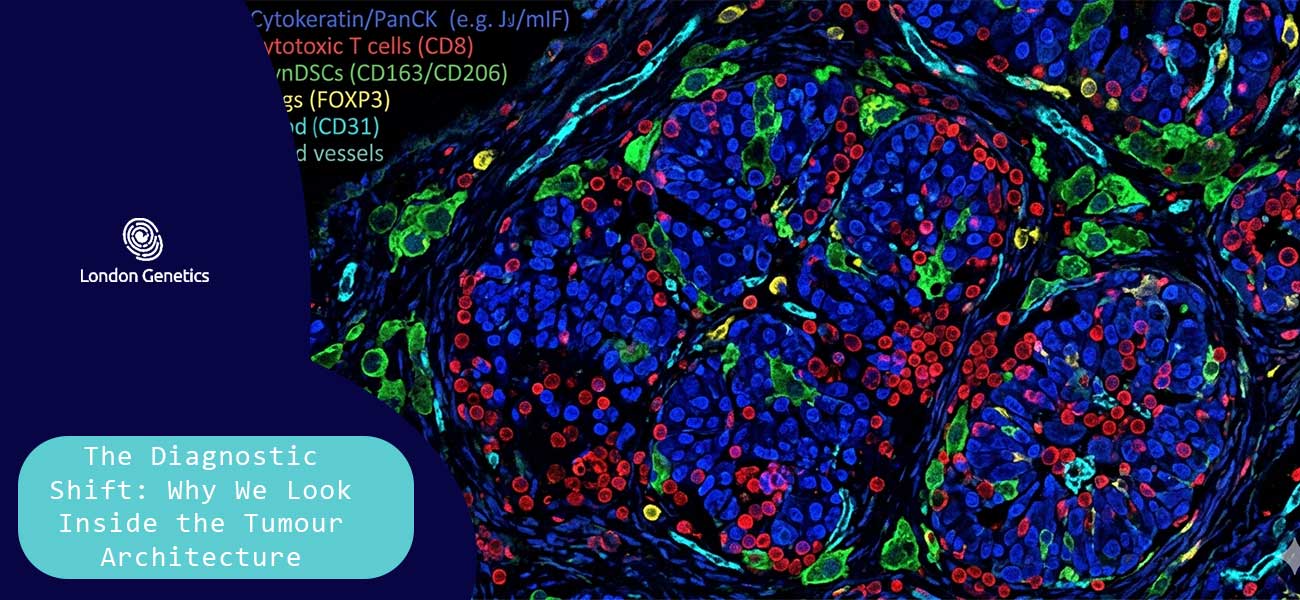
For decades, conventional oncology relied almost exclusively on anatomical landmarks—evaluating the size, location, and staging of a growth through standard scans and physical biopsies. However, two identical-looking masses under a microscope can behave in entirely opposing ways, responding differently to identical treatments. This clinical variation occurs because the true driver of a malignancy is hidden deeper within its unique molecular profile. To achieve diagnostic precision, modern medicine has undergone a profound shift, moving away from generalized assumptions and transitioning toward advanced somatic genomic testing to read the exact chemical blueprint of the altered tissue.
The biological necessity for this localized diagnostic approach comes down to where these genetic errors physically reside. Because somatic mutations are entirely acquired throughout life, they are locked strictly within the expanding mass. They do not exist in the patient’s healthy white blood cells, skin cells, or saliva.
Consequently, traditional hereditary blood tests or standard liquid screenings are fundamentally incapable of detecting them. If a clinical team only analyses a patient’s healthy blood sample, these critical, tissue-specific alterations remain completely invisible. To uncover the true drivers of the disease, specialists must bypass healthy tissues entirely and perform a direct, comprehensive tumour tissue analysis.
┌────────────────────────────────────────────────────────────────────────┐
│                    THE DIAGNOSTIC DETECTION BARRIER                    │
├───────────────────────────────────┬────────────────────────────────────┤
│  STANDARD BLOOD/SALIVA TEST       │  SOMATIC GENOMIC TESTING           │
├───────────────────────────────────┼────────────────────────────────────┤
│ • Scans the inherited germline    │ • Scans the isolated tumour tissue │
│ • Misses local acquired errors    │ • Identifies specific active mutations│
│ • Blind to somatic alterations    │ • Captures real-time drivers       │
│ • Result: INCOMPLETE PROFILE      │ • Result: ABSOLUTE CLARITY         │
└───────────────────────────────────┴────────────────────────────────────┘
When a pathologist extracts a sample and subjects it to somatic genomic testing, the entire cellular landscape is illuminated. This advanced sequencing method acts as a high-definition scanner, mapping out the precise layout of the mass to isolate the exact somatic mutations that caused the cells to lose control.
By prioritizing this deep tumour tissue analysis, healthcare providers no longer have to guess which pathways are fueling the disease. Instead, utilizing somatic genomic testing allows clinicians to pinpoint the exact molecular vulnerabilities of the target area, shifting the medical approach from a retrospective analysis of symptoms to a proactive, highly targeted intervention.
Conclusion: Tailoring Targeted Therapies to Your Molecular Blueprint
The ultimate value of identifying somatic mutations does not lie in simply cataloguing cellular errors; it lies in the profound therapeutic power that this data unlocks. In the era of modern precision medicine, cancer management has evolved past treating a disease based solely on its anatomical location, moving away from generic treatments toward a highly sophisticated, bespoke management plan.
The journey through advanced genomic science should never be walked alone. At London Genetics, we are dedicated to bridging the gap between theoretical diagnosis and absolute clinical control, replacing standard broad-spectrum protocols with a clear, confident, and life-saving blueprint tailored entirely to your body’s true molecular reality.
Advanced Tissue Profiling:
Running comprehensive somatic genomic testing to read your unique tissue architecture and pinpoint the exact driving genetic mutations altering your cells.
Clinical Data Contextualisation:
Translating complex tumour profiles and somatic mutations alongside your active medical history during a highly specialised genomic consultation session.
Bespoke Targeted Architecture:
Deploying engineered, patient-specific targeted therapies that function like heat-seeking missiles, blocking abnormal proteins while sparing surrounding healthy cells.
Proactive Genetic Pathway:
Bypassing generic, broad-spectrum interventions by isolating specific molecular pathways and variants such as EGFR or BRAF to disrupt the growth of the disease.
Definitive Health Control:
Stepping out of standard clinical protocols and general statistical tracking to take absolute biological control equipped with elite genetic precision.